
Step-by-Step
Mohs Surgery Process
Mohs surgery follows a carefully structured process designed to remove skin cancer with precision. This page outlines each step of the Mohs procedure, from preparation through tissue analysis and completion, so patients know what to expect on the day of treatment.
Mohs surgery is a specialized form of dermatologic surgery designed to remove certain types of skin cancer with a high level of precision. Understanding each step of the process can help reduce uncertainty and set clear expectations before treatment. The procedure is performed in stages, allowing the Mohs surgeon to remove cancerous tissue while preserving as much healthy skin as possible.
This page walks through the Mohs surgery process from start to finish, explaining what happens on the day of surgery, how tissue is examined, and what to expect between each step. The goal is to provide clear, patient-focused information to help you feel informed and prepared as you move forward with skin cancer removal.
Overview of the Mohs Surgery Process
Mohs surgery follows a structured, step-by-step approach that allows for careful removal of skin cancer while protecting surrounding healthy tissue. Each stage of the procedure is guided by real-time examination of the tissue, helping ensure that cancer cells are fully removed before reconstruction begins. This method is especially valuable for areas where preserving normal skin is important.
What Makes Mohs Surgery Unique
Mohs surgery is different from other skin cancer treatments because the Mohs surgeon examines the entire surgical margin during the procedure. After each layer of tissue is removed, it is processed and evaluated under a microscope on-site. This allows the surgeon to precisely identify and remove remaining cancer cells while minimizing unnecessary removal of healthy skin.
Goals of the Step-by-Step Approach
The step-by-step process is designed to balance complete skin cancer removal with optimal cosmetic and functional outcomes.
Key goals include:
Removing all cancerous cells with accuracy
Preserving as much healthy tissue as possible
Reducing the risk of cancer recurrence
Supporting effective wound healing and reconstruction
This careful approach makes Mohs surgery a trusted option for treating certain basal cell carcinoma and squamous cell carcinoma cases.
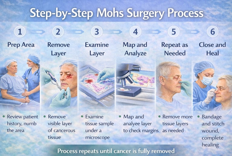
Step 1: Preparing the Treatment Area
The first step of Mohs surgery focuses on careful preparation to ensure accuracy, comfort, and safety. This stage sets the foundation for precise skin cancer removal and helps the Mohs surgeon clearly identify the area requiring treatment.
Confirming the Surgical Site
Before the procedure begins, the Mohs surgeon confirms the exact location of the skin cancer. This may involve reviewing biopsy results, examining the area, and discussing the site with the patient. Confirming the surgical site helps ensure that the correct area is treated and that the procedure is performed with precision.
Local Anesthesia
Once the treatment area is confirmed, local anesthesia is administered to numb the skin. This helps keep the patient comfortable during the procedure while allowing them to remain awake. Most patients experience minimal discomfort after the anesthetic takes effect, and general anesthesia is not typically required for Mohs surgery.
Step 2: Removing the First Layer of Skin Cancer
After the treatment area is numbed, the Mohs surgeon begins removing the first thin layer of cancerous tissue. This step is performed with great precision and is designed to remove visible cancer while sparing as much healthy skin as possible.
Precise Tissue Removal
The Mohs surgeon carefully removes a narrow layer of tissue from the affected area. Unlike standard excisions, Mohs surgery removes tissue in a controlled and targeted manner. This approach helps ensure effective skin cancer removal while minimizing the size of the surgical wound.
Mapping the Tissue
Once the tissue is removed, it is carefully mapped and labeled. This detailed map acts as a guide, showing exactly where each piece of tissue came from on the skin. Tissue mapping allows the Mohs surgeon to identify the precise location of any remaining cancer cells during microscopic examination, guiding subsequent steps if additional tissue removal is needed.
Step 3: Microscopic Examination of the Tissue
Following removal of the tissue layer, the sample is analyzed to determine whether any skin cancer cells remain. This step is central to Mohs surgery and allows the Mohs surgeon to evaluate the entire surgical margin with a high level of accuracy.
Processing the Tissue in the On-Site Lab
The removed tissue is taken to an on-site laboratory, where it is carefully processed and prepared for microscopic examination. The tissue is frozen, thinly sectioned, and placed onto microscope slides. This preparation allows the surgeon to examine the edges and underside of the tissue in detail.
Examining the Margins Under the Microscope
The Mohs surgeon examines the processed tissue under a microscope to look for remaining cancer cells along the margins. Because the entire margin is evaluated, this step helps identify even small areas where cancer may still be present. If cancer cells are found, their exact location is marked on the tissue map, guiding the next stage of targeted skin cancer removal.
Step 4: Determining Whether Cancer Remains
Once the tissue has been examined under the microscope, the Mohs surgeon determines whether any cancer cells are still present. This decision guides the next step in the procedure and helps ensure complete skin cancer removal.
Identifying Residual Cancer Cells
During microscopic examination, the Mohs surgeon looks for any remaining basal cell carcinoma or squamous cell carcinoma at the edges of the removed tissue. Because the tissue is carefully mapped, the surgeon can identify the exact location where residual cancer cells may remain. This precision helps limit additional tissue removal to only the affected areas.
Deciding if Another Stage Is Needed
If cancer cells are detected, another stage of Mohs surgery is performed to remove an additional thin layer of tissue from the specific area involved. If no cancer cells are found, the removal portion of the procedure is complete. This step-by-step process continues only as needed, helping preserve healthy skin while achieving effective skin cancer removal.
Step 5: Additional Stages (If Necessary)
Some skin cancers extend beyond what is visible on the surface. When this occurs, additional stages of Mohs surgery are performed to ensure that all cancer cells are removed while preserving healthy tissue.
Why Multiple Stages May Be Required
Skin cancer can grow with irregular or microscopic extensions that are not apparent during the initial removal. Basal cell carcinoma and squamous cell carcinoma may spread unevenly beneath the skin, especially in previously treated or long-standing lesions. Multiple stages allow the Mohs surgeon to identify and remove these hidden cancer cells with precision.
How Additional Layers Are Removed
If residual cancer is found, the Mohs surgeon removes another thin layer of tissue only from the area where cancer remains. This targeted approach avoids unnecessary removal of healthy skin. Each additional layer is processed and examined in the same careful manner until no cancer cells are detected.
Step 6: Confirming Complete Cancer Removal
The final stage of the cancer removal portion of Mohs surgery occurs once microscopic examination shows that no cancer cells remain. This confirmation is a key advantage of Mohs surgery and provides clarity before moving on to wound care or reconstruction.
Clear Margins Achieved
Clear margins mean that the edges of the removed tissue are free of cancer cells when examined under the microscope. By evaluating the entire margin, the Mohs surgeon can confirm that basal cell carcinoma or squamous cell carcinoma has been fully removed from the treated area. This careful verification helps reduce the risk of the cancer returning in the same location.
Ending the Cancer Removal Phase
Once clear margins are confirmed, no further tissue removal is needed. At this point, the skin cancer removal phase of Mohs surgery is complete. The procedure then transitions to discussing wound healing options, which may include allowing the area to heal naturally or performing surgical repair, depending on the size and location of the wound.
Step 7: Wound Repair & Reconstruction
After complete cancer removal is confirmed, attention turns to repairing the surgical site. The goal of wound repair and reconstruction is to support proper healing while preserving function and appearance whenever possible.
Deciding on the Best Repair Option
The most appropriate repair method depends on several factors, including the size, depth, and location of the wound. Options may include allowing the area to heal naturally, closing the wound with stitches, or using specialized reconstructive techniques. The Mohs surgeon considers both medical and cosmetic factors when selecting the repair approach.
Transition to Reconstruction
Once a repair plan is determined, reconstruction may be performed immediately following the cancer removal phase. In many cases, the Mohs surgeon completes both the removal and reconstruction on the same day. This seamless transition helps streamline care and allows patients to move forward with healing after their dermatologic surgery.
Why the Step-by-Step Process Improves Outcomes
Mohs surgery is designed around careful, staged skin cancer removal with immediate microscopic confirmation. This step-by-step method supports both medical effectiveness and thoughtful preservation of healthy skin, which can be especially important in visible or functionally sensitive areas.
Precision & Tissue Preservation
Because Mohs surgery removes tissue in thin layers and checks each layer under the microscope, treatment can be directed only where cancer cells remain. This precision helps the Mohs surgeon remove basal cell carcinoma or squamous cell carcinoma while sparing as much normal skin as possible. Preserving healthy tissue can support better wound repair, reconstruction, and overall healing.
Highest Cure Rates
Mohs surgery is widely recognized for offering very high cure rates for many basal cell carcinoma and squamous cell carcinoma cases, particularly when tumors are high risk or have returned after prior treatment. The ability to examine the surgical margins during the procedure helps confirm complete cancer removal before reconstruction begins. While no treatment can guarantee a specific result for every patient, this staged approach is one reason Mohs surgery is often recommended for certain skin cancers.
Frequently Asked Questions About the Mohs Process
Common Process-Related Questions
How long does Mohs surgery take?
Mohs surgery is performed in stages, so the total time can vary. Most procedures take several hours, including waiting periods while tissue is examined. Patients are usually advised to plan for the majority of the day.
Will I be awake during the procedure?
Yes. Mohs surgery is performed using local anesthesia, so patients remain awake but comfortable throughout the procedure.
Why is there waiting time between stages?
Waiting periods allow the removed tissue to be processed and examined under a microscope in the on-site lab. This step ensures that any remaining cancer cells are identified before additional tissue is removed.
How many stages of Mohs surgery will I need?
The number of stages varies depending on how far the cancer extends beneath the skin. Some cancers are removed in a single stage, while others require multiple stages to achieve clear margins.
Is reconstruction done the same day?
In many cases, wound repair or reconstruction is performed on the same day once complete cancer removal is confirmed. The timing and method depend on the size and location of the surgical site.


Request a Mohs Consultation
Copyright © 2026 | Savannah Mohs Surgery | All Rights Reserved.
Privacy Policy | Terms and Conditions | Accessibility
Monday: 8:00am - 5:00pm
Tuesday: 8:00am - 5:00pm
Wednesday: 8:00am - 5:00pm
Thursday: 8:00am - 5:00pm
Friday: 8:00am - 5:00pm
Office Hours
Learn More
Reach Out
Resources
Address
Request Consultation
Call Now
(704) 858-6585
Savannah Mohs Surgery
Serving Savannah, GA and Surrounding areas.

